Wednesday, September 16, 2015

Rain, Rain gauge And Ganesha Chaturthi


When we put up a fabricated structure to cover a part of our compound space, the fabricator had to leave spaces here and there to allow the electric wires, telephone cables and TV cables to pass through.  Rain water was flowing through these gaps and to prevent that we did some ‘Jugaad’ and managed to close most of them.  Still, one spot remained open and drops of 
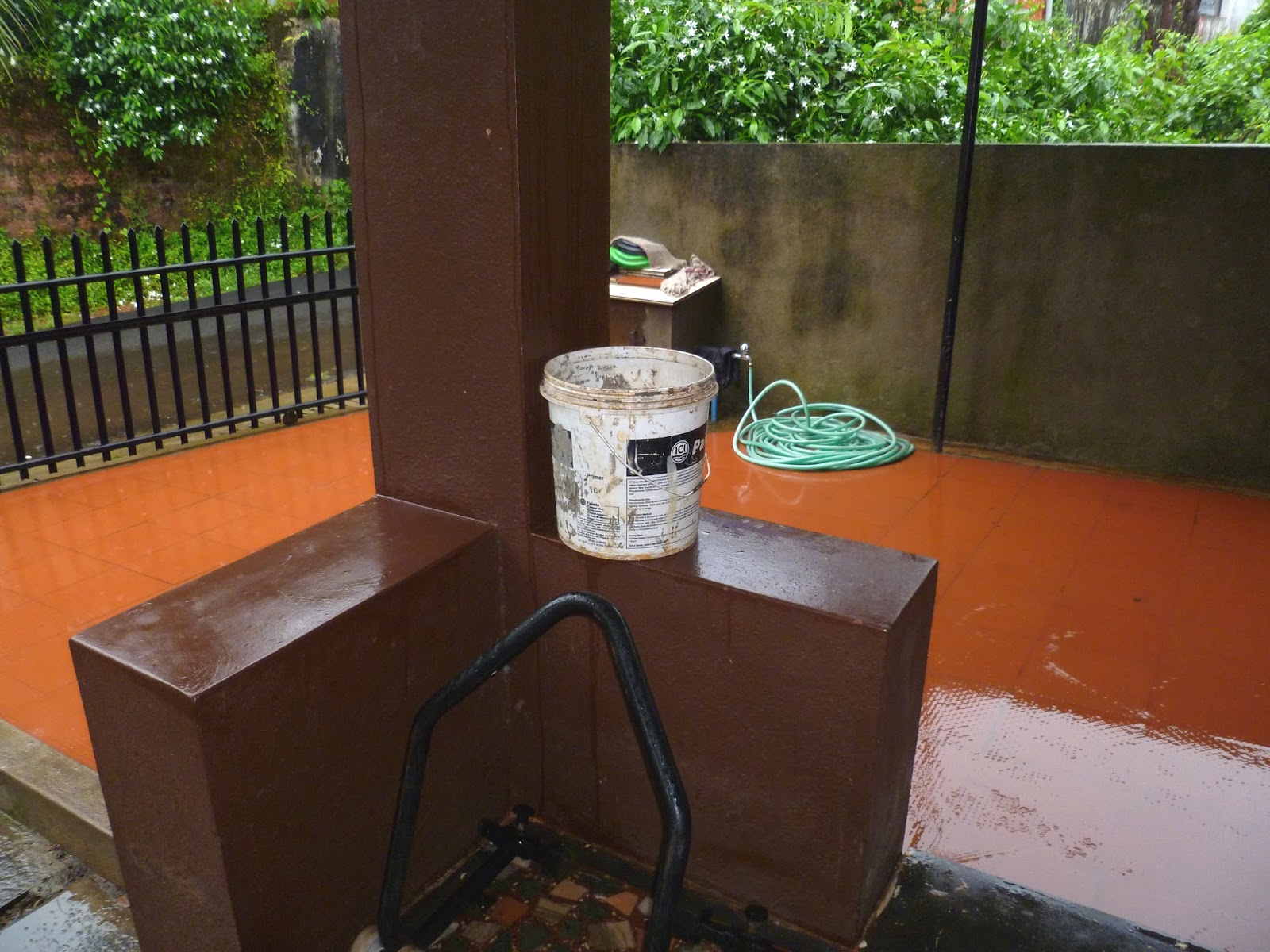
rain fell into our portico. I managed it with another simpler ‘Jugaad’, which, in this case, is a bucket placed to catch these leaking drops. This bucket is my rain gauge. I empty it every morning and from the amount of water collected in this bucket I can measure the rain fall on my house as accurately as the meteorological department.  For most of this rainy season the bucket remained dry.  A week back the weather department announced that the monsoon has retreated and that the rain fall this season is deficient 25%. Tomorrow is Ganesha Chaturthi and I have hardly seen a rain free ‘chaturthi’ here. I thought this may be one.


Ganesha chaturthi is the most widely celebrated festival in Goa. It is a big issue here. From all points of view - religious, social, commercial and political. The bazaar starts buzzing with ‘chouti’ related activities at least a week or two before the festival.  These activities peak two days preceding ‘chouthi.’ There is a tradition of decorating the ‘mantap’ (‘matoli’ as it is locally known) with locally grown vegetables as also wild fruits, flowers and leaves.  In Ponda, the main bazaar road is closed for traffic two days before ‘chouti’ and the street is fully occupied by people selling and buying these things. It is called the ‘matoli bazaar’.  The bazaar began yesterday and it has been raining without break since then.  My rain gauge has recorded 3 cms of rain since yesterday. I hear that people are having a tough time shopping for the festival. The rain, which should have been welcome, is being cursed.

Since I hardly have anything else to do, we have finished our purchases days ahead of ‘chaturthi’ and I am now sitting waiting for my wife to finish her preparations and begin the Gowri pooja. I am the officiating priest and am under strict orders not to leave the room before I finish my task. I am sitting  snug enjoying the rain and using the time to type whatever occurred to my mind.


Goa badly needed rain and I heard that some people performed a ‘Yagna’ at the ‘Brahma’ temple last week.
They should have prayed for rains to begin AFTER Ganesha Chaturthi.


Friday, September 4, 2015

Teaching Dentistry - My Patient Education Programme


Most of my patients think that the ‘gum’ is a block of flesh and the teeth remain stuck in it somehow or the other. A tooth starts paining when a cavity goes deeper and touches the ‘flesh’ below.  A ‘filling’ is something similar to what a mason does. The mason mixes some cement and plugs a hole in the wall and the dentist mixes some cement (many may even be thinking that it is the same cement) and plugs a hole in the tooth. A ‘fixed’ tooth is somehow glued to the gum or tied to other teeth using some wires.  All these may sound idiotic to a dentist but I understand. My knowledge of an electric transformer or a petrol engine or a TV tube would be equally bad if not worse.  I should be least bothered about what my patients think as long as they get the treatment done and pay my fee. But I have a defect. Rather, I have many defects and one of them is trying to make my patients understand what the treatment involves. I spend considerable time and use a variety of teaching aids - a skull and mandible, plaster of Paris working models, plastic exhibition models, extracted teeth and on the spot drawings to achieve my goal. I give detailed explanations about impactions, dentures, RCTs, bridges, braces and what not and at the end, usually receive a bored expression and a blank stare for my efforts. But I persist.

My teaching aids. Ever seen a roadside dentist? His spread on the footpath to advertise his profession is exactly like this.

Recently we had an unusual case in the clinic where in a boy came in for braces to correct his alignment and the x ray showed an impacted canine (unerupted eye tooth stuck in the jaw bone) in the upper jaw. Since it could give rise to complications later in life and since it was mandatory to get rid of it before proceeding with orthodontic treatment (Braces), we decided to remove it. It was a big tooth, and it would have left a big hole in the jaw after removal. It would have taken a long time to close up. Hence, our young surgeon Dr Saurabh suggested that we remove a piece of bone from the chin region (lower jaw) and pack it (graft) in the hole after removing the canine tooth in the upper jaw. The procedure which can be called “extraction of an impacted upper canine followed by autogenous bone graft” was planned.

This is an X Ray of all the teeth in the mouth, known as orthophantamograph. OPG for short. Tooth C is the central incisor, L is the lateral incisor M  is the milk tooth and Ca is the impacted canine. In the normal course this Ca should have pushed the milk tooth out and taken its place. But it changed course, came between the two incisors creating trouble and forcing extraction (removal)


This is a CBCT (Cone Beam Computed Tomography) of the area marked in the other x ray. Something like a CT scan. Gives much more information than an OPG and helps in accurately locating the structures - in this case the canine. Makes the job a little easy for the surgeon.


Opened up the place where the canine is expected to be hiding. The 'gum' is not a mass of flesh. It is a tissue layer about 2 mm thick and covers the jaw bone inside the mouth. Two cuts are made on either side of the operation area and the gum is peeled and pushed up exposing the jaw bone.

The jaw bone is scraped off with a surgical drill exposing the impacted tooth lying underneath. What is seen is about one third of the tooth called the crown. The root, which is two thirds, is not exposed and is inside the bone.


The exposed part, the crown, is cut, separated from the root and taken out


The root as seen after removing the crown.
Root is pulled down into the space crated after removing the crown and is taken out.
The hole in the jaw after the removal of the tooth


What you are seeing here is the cut made to reach the chin bone. The cut is behind the lower lip in front of the lower front teeth 

The rectangular 'window' seen above the retractor is the place from which fragments of bone are removed for grafting.

Bone fragments (graft) placed in the cavity earlier occupied by the tooth

The cut behind the lower lip is sutured.

The cut made for removal of the impacted canine is sutured.
As a surgery it is not something great but it was the first time that such a procedure was being done in my clinic. I was excited as well as apprehensive. Cutting open the upper jaw was compulsory. Cutting the lower jaw was optional. Patients get jittery the moment a knife comes into picture. We were suggesting an extra cut which was not a necessity though beneficial to the patient.  The parents seemed to understand.  Saurabh as usual was confident. We did the extraction and graft. It took two hours. The boy bore it bravely. The parents were scared to look at the procedure and spent the time worrying, watching TV and napping.  It is a week since we did the case and the boy is fine. I had taken pictures at different stages so that I could explain the procedure to his parents and since I had the pictures, thought of posting them here. There is no reason why anyone should be interested in the gory details of my profession but you don’t know. This is an era where people are interested in gory details of everything. Whatsapp brings you all the gory details of an accident or suicide and TOI is full of a murder which took place three years back. This is far better. If you happen to be one of the interested, and have gone through the post, please let me know if you could make out what was done. That would help me rate my capabilities as a teacher, and if so mandated, put an end to my stupidity of trying to teach dentistry to my patients.